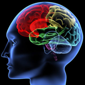
 After having identified REM sleep as the privileged moment of the dream, scientists wanted of course to know more.
How, precisely, does this phase of REM sleep occur? How and where does the dream originate in our brain? Many elements are still missing, but the teams of sleep specialists and neuroscientists have allowed science to move forward on the issue.
Brain imaging and inconsistent dreams
First observation: during the REM phase, the primary visual areas are deactivated, that is to say that the sense organs do not transmit information to the brain.
However, the brain continues to form images in other more sophisticated visual areas: it seems that it functions in autarky and does not need external images to function. In the same way as when a person closes his eyes and makes an effort of imagination, he can easily visualize the landscape of his dreams, for example.
Except that in the case of the dream, it was not his will that made an effort to recall this image. What remains to be investigated is how this image was created in the brain.
What we do know, however, is that the prefrontal lobe, which partly manages the consistency of the information we receive, is at rest during REM sleep. So there is no more censorship and our brain can no longer tell the difference between what is true, what is not, what is coherent or not.
Hence the difficulty in telling our dreams on waking: they often have neither tail nor head and no logical frame.
Birth of dreams: an experience on cats
Neuroscientists wanted to understand what was causing this altered state of the brain and to verify where exactly REM sleep was born: is there a dream center? These researchers therefore conducted an exciting experiment on cats. We already knew that the cortex was not essential for REM sleep.
The teams therefore concentrated on the brainstem and gradually removed small parts, notably the hypothalamus and the pituitary gland, two structures which are nevertheless very important in regulating moods. Each time they cut a part of the brainstem, they found that cats continued to experience very regular REM sleep periods, neither the duration nor the frequency of which were shortened.
Second stage of the experiment: there remains only the bridge, a structure which makes the link between the brain and the spinal cord. The researchers decided to practice progressive bilateral lesions on this element.
Very quickly, they realized that, this time, the mutilated cats no longer had REM sleep and meaningful dreams at all: it would therefore seem that it originates in this bridge, more precisely at the level of its dorsolateral part.
The role of neurotransmitters
These data were confirmed by histochemical experiments which made it possible to show the accumulation of enzymes, responsible for the destruction of monoamine oxidases at the level of a very close structure, the cerulean complex.
This explains in particular that when a person has been deprived for a certain period of REM sleep, the duration of the dream, each night, increases: the enzymes seem to regulate the release of these monoamines over the long term.
This theory is now confirmed: thus, when monoamine oxidase inhibitors are injected into an individual, the phases of REM sleep (and therefore the dream in its strict version) disappear.
It remains to be seen how this monoamine oxidase then acts on the brain. It makes it possible to inactivate certain neurotransmitters of the monoamine class, such as norepinephrine, serotonin or dopamine. All these neurotransmitters play, among other things, a role in the regulation of mood and sleep.
Monoamine oxidase therefore has an obvious indirect effect on the different phases of sleep. Later, a team of Swedish researchers from the Karolinska Institute helped define a topography of the circuits of these neurotransmitters. Systems that would also be able to control each other and that researchers are still far from finished deciphering.
We know in particular that serotonin is necessary for falling asleep and keeping the body in a state of sleep. In the morning, serotonin inhibitors take effect, its concentration in the body decreases and we wake up.
The two systems are linked: if we deactivate the noradrenergic neurons, then the serotonergic neurons activate and the amount of daily sleep increases considerably, as does the quantity of dreams.
As for the REM sleep phase in particular, teams have identified in the brainstem the structures responsible for its activation. It is a set of SP-on neurons, active during REM sleep.
These neurons have their reverse equivalent, the SP-off neurons, which work thanks to aminergic neurotransmitters (noradrenaline, serotonin, histamine): they prevent the SP-on neurons from functioning. It is only when their action stops that they can “light up” and trigger REM sleep.
Another peculiarity of REM sleep, metabolic this time: experience has shown that the energy expenditure of the organism in glucose and oxygen was the same as during waking: better to have eaten well the night before if we want to have enough energy to dream! Spending is significantly slowed down during slow sleep.
Enzymes are therefore responsible for the ability to dream; the more there are, the less a person dreams. The mechanism of the latter is therefore very biochemical, even metabolic.